Fig. 4.
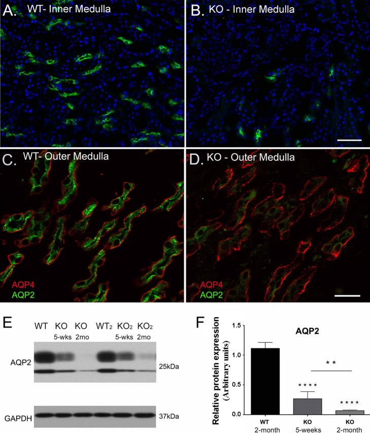
Deletion of β1-integrin in principal cells causes a significant decrease in AQP2 expression in certain tubules. AQP2 (green) shows normal expression and distribution in the inner (A) and outer medulla (C) of WT mice. AQP2 immunostaining is significantly reduced in the β-1f/fAQP2-Cre+ (KO) medulla (green in B and D), while AQP4 immunostaining is preserved in most KO collecting ducts (red in D). Scale bar = 100 μm for A and B and 40 μm for C and D. E: the loss of AQP2 expression in KO mouse collecting ducts was confirmed by Western blot analysis. F: a bar graph representing the total AQP2 protein expression levels from Western blotting results. There was a significant decrease in AQP2 protein in 5 wk old KO animals, which continues to decrease significantly to almost total loss by ~2 mo of age. Bar values represent means ± SE. Statistical analysis was performed with one-way ANOVA. **P < 0.01 and ****P < 0.0001; n ≥ 3.

