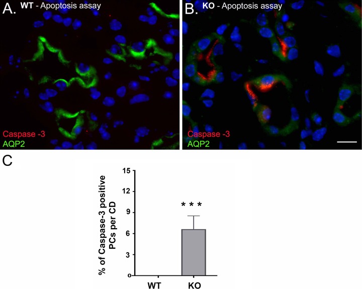
Fig. 5.
Confocal microscopy images of 5-wk-old β-1f/fAQP2-Cre+ (KO) and wild type (WT) mouse renal medulla. A: there is no active/cleaved caspase-3 expression (red) in WT medulla. B: active/cleaved caspase was detected in KO collecting duct (CD) principal cells (PCs). C: bar graphs showing the percentage of active/cleaved caspase-3 positive PCs per CD. Scale bar = 20 μm. Bar values represent means ± SE. Statistical analysis was performed with Student’s t-test. ***P < 0.001; n ≥ 3.

