Fig. 7.
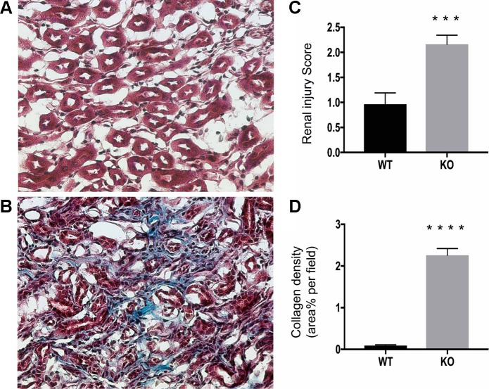
Diffuse interstitial fibrosis in β-1f/fAQP2-Cre+ (KO) kidneys by trichrome stain (Masson). A: representative photomicrograph of a Masson-stained kidney section of a 2-mo-old WT mouse showing no collagen deposition in the outer medulla. B: the degree of collagenous material in blue, cytoplasm and muscle fibers in red and nuclei in black. Collagen deposition was significantly higher in KO mouse outer medulla than in WT littermates. C: histological injury score for medulla samples stained with Hematoxylin and eosin. Injury scale: 1.0 - 3.0, with 1.0 being normal. D: quantification of collagen deposition in Masson-stained kidney section. Bar values represent means ± SE. Statistical analysis was performed with Student’s t-test. *P < 0.05, **P < 0.01, ***P < 0.001, and ****P < 0.0001; n ≥ 3.

