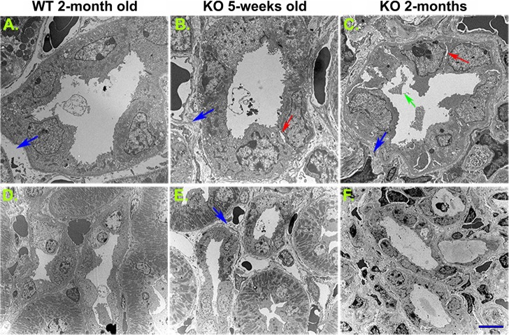
Fig. 8.
Collecting duct tubular injury and peritubular fibrosis as revealed by transmission electron microscopy. Compared with the 2-mo-old wild-type animals (A and D), a disruption of intercellular junctions was detected in 5 wk old and more obviously 2 mo-old β-1f/fAQP2-Cre+ (KO) animals (arrows in B and C). Collecting duct cell debris was clearly seen in the lumen of 2-mo-old KO mouse collecting ducts (green arrow in C). Significant peritubular extracellular matrix deposition and interstitial fibrosis were visible in 5-wk-old (B and E) and more evident in 2-mo-old KO mice (C and F). Scale bar = 4 μm (A–C) and 10 μm (D–F). Blue arrows indicate basolateral extracellular matrix space. Red arrows indicate collecting duct cell detachment from neighboring cells. The green arrow indicates cell debris in the collecting duct lumen.

