Fig. 6.
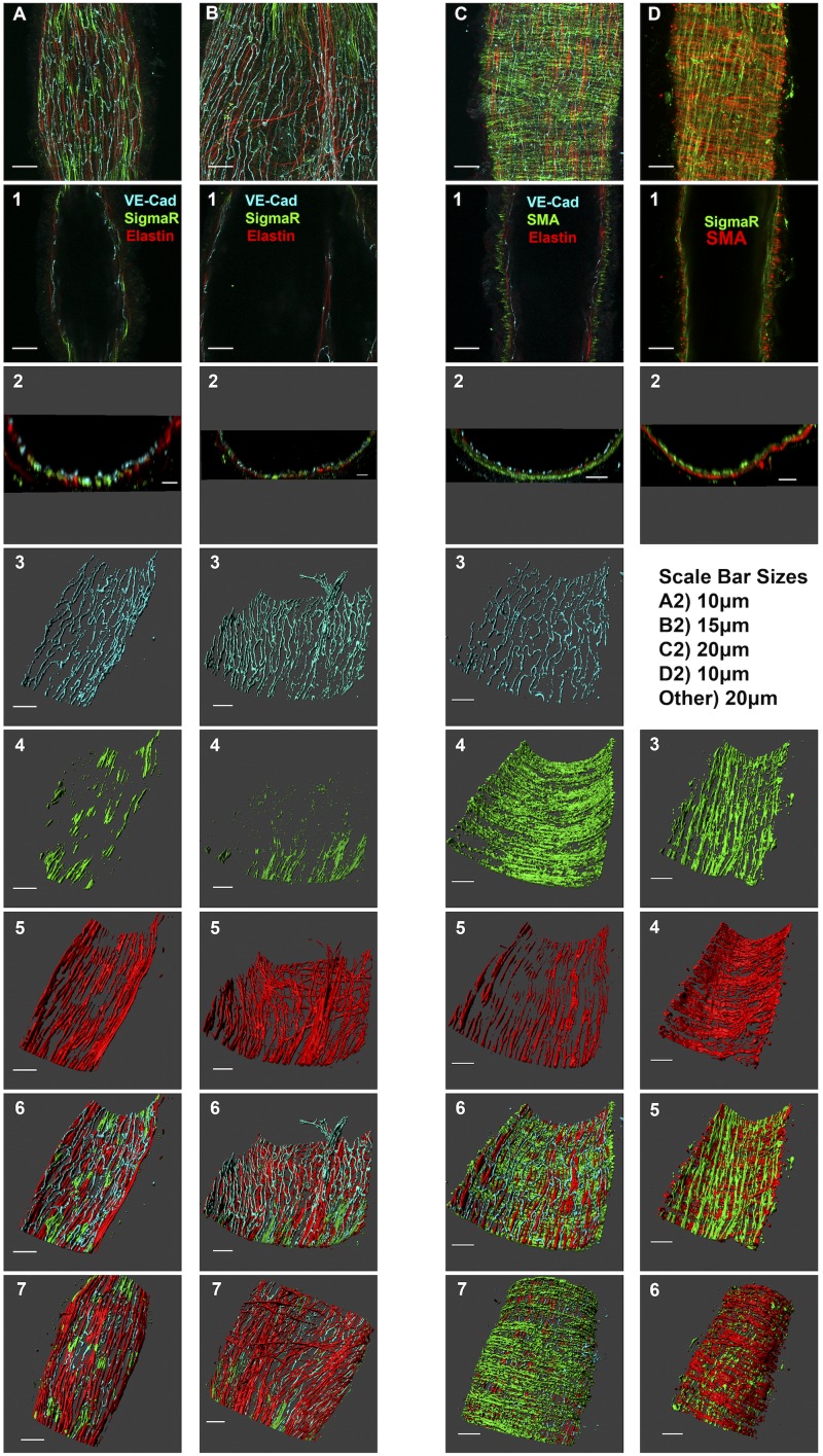
Localization of the σ1-receptor in isolated rat collecting lymphatic vessels. A and B: images of two different collecting lymphatic vessels that were labeled for vascular-endothelial (VE)-cadherin, the σ1-receptor, and elastin. C: labeling for VE-cadherin, elastin, and smooth muscle actin. D: σ1-receptor and smooth muscle actin labeling. Top images show a maximum-intensity z-projection obtained with a ×60 objective. A−D,1: representative confocal slices about halfway through the z-stack, with the target proteins denoted by the colors in the label. A−D,2: orthogonal projections (5 µm thickness) representing a cross-sectional view of the vessel. Images 3–5 in A–C and images 3 and 4 in D show three-dimensional representations of individual channels, with the luminal side of the vessel facing upward. Image 6 in A–C and image 5 in D show three-dimensional composites of all channels, with the luminal side of the vessel facing upward. Image 7 in A–C and image 6 in D show three-dimensional composites of all channels but with the abluminal side of the vessel facing upward. Each labeling scheme is representative of 3 vessels from 3 rats each.

