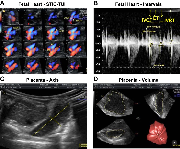
Fig. 1.
Spatiotemporal image correlation (STIC) and tomographic ultrasound imaging (TUI) of fetal heart (A), Doppler images of cardiac intervals (B), and 2-dimensional (C) and 3-dimensional (D) images of placenta. Four-dimensional echocardiography with STIC-TUI, along with color Doppler, was used to evaluate structural features of the fetal heart. Doppler velocity waveforms of right and left outflow tracts were obtained in the 4-chamber view to measure time intervals during the cardiac cycle. 2-Dimensional placental images were obtained using Doppler ultrasound for short and long axes of the placenta. 3-Dimensional placental images were obtained using virtual organ computer-aided analysis (VOCAL) software to determine placental volume. ET, ejection time; IVCT, intraventricular contraction time; IVRT, intraventricular relaxation time.

