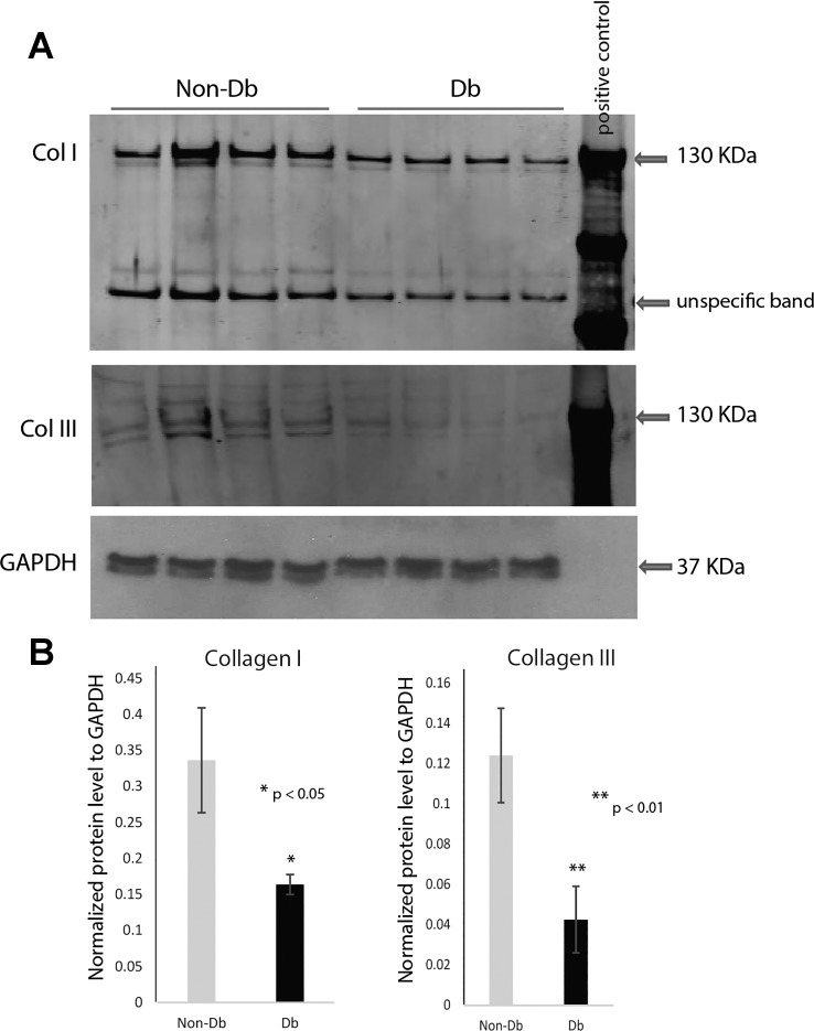
Fig. 1.
Western blot analysis demonstrating decreased collagen content of diabetic wounds. A: Western blot analysis of collagen I and III protein content and loading control GAPDH in nondiabetic (Non-Db, Db/+) (n = 4) and diabetic (Db, Db/Db) (n = 4) wounds 7 days after injury (top). Equal amounts of protein samples were loaded to each lanes: 4 for nondiabetic wounds, 4 for diabetic wounds, together with 1 lane for positive control. The positive control was loaded with standardized either collagen I or collagen III protein. B: normalized collagen I and III content was calculated after normalizing with internal loading control (bottom) using Image J analysis. Results are presented as means ± SD; *P value <0.05 considered statistically significant by Student’s t-test. Comparison was performed between Db/Db wounds and Db/+ wounds. Diabetic wounds showed significantly lower level on collagen I or collagen III expression.

