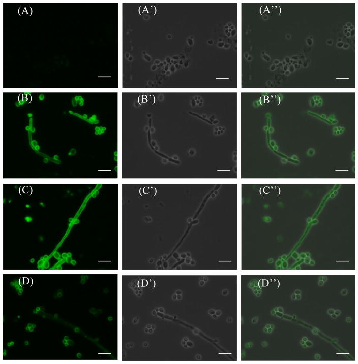
Figure 4.
IF staining results of the heat-killed C. albicans cells using sera obtained from mice (A) without vaccine treatment (negative control) or immunized with glycoconjugates (B) 1a, (C) 2a, and (D) 3a. Sub-panels A′ through D′ show the corresponding bright field images, and sub-panels APrime; through DPrime; show the merged images. The length of the white bar is 10 μm.

