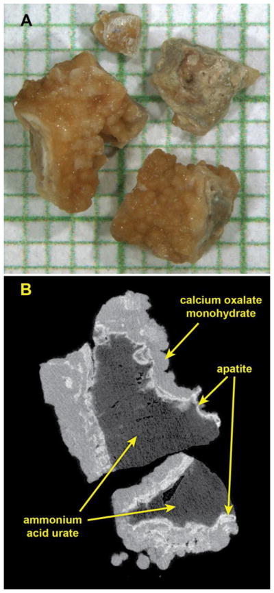
Figure 1.

Micro CT characterization of a typical specimen of mixed mineral in this study. A: Photo of stone fragments on mm-grid paper. B: Micro CT image slice through two of the fragments. X-ray attenuation values indicated that these stone fragments contained a core of urate or uric acid (dark gray, determined by infrared spectroscopy to be urate), surrounded by a shell composed of calcium phosphate in the form of apatite (bright white regions) and calcium oxalate monohydrate (gray regions). This specimen was judged to be sufficiently uniform among fragments that it could be divided randomly and submitted for both clinical FT-IR and CA analysis.
