
Fig. 4.
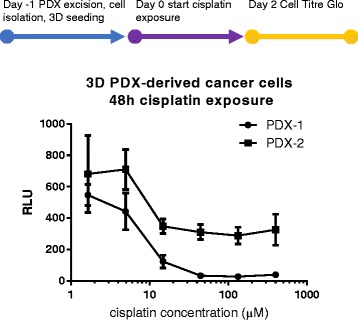
Cisplatin exposure of PDX-derived human breast cancer cells in 3D microfluidic culture. Human cancer cells from two different breast cancer PDX avatars were isolated and seeded in 3D in the OrganoPlate® 1 day prior to 48 h cisplatin exposure. Culture viability was quantified using the luminescent CellTiter-Glo® cell viability assay. IC50 were determined based on nonlinear fit of the dose response range as 8,1 μM and 14,8 μM for PDX-1 and PDX-2 respectively