Fig. 4.

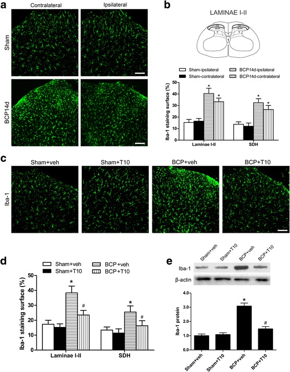
Effect of intrathecal administration T10 on microglial activation in the spinal dorsal horn (SDH) of BCP rats on POD 14. a Immunostaining of spinal sections showed the BCP-induced change of microglia marker, Iba-1, in the SDH. b Quantitative analysis of the percentage of Iba-1 immunostaining surface in the SDH showed the BCP-induced change (n = 4). c Immunostaining of Iba-1 in the ipsilateral SDH. d The percentage of Iba-1 immunostaining surface in the laminae I–II and whole SDH on the ipsilateral side. e Western blot analysis of the protein level of Iba-1 in the ipsilateral SDH, relative to that of sham + vehicle group (n = 5). Scale bar 100 μm. *p < 0.05 vs. sham + vehicle group; # p < 0.05 vs. BCP + vehicle group
