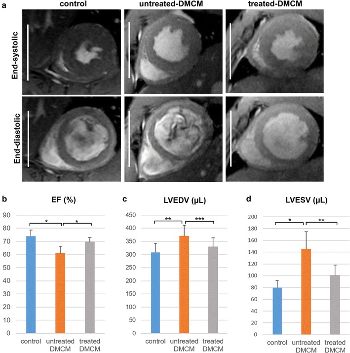
Fig. 4.

Cardiac magnetic resonance imaging reveals preserved left ventricular geometry following cell sheet therapy. a Representative cardiac MRI images at end-systolic and end-diastolic phase for each experimental group. b–d In the cell sheet treatment group, left ventricular ejection fraction (b) was significantly improved and both left ventricular end-diastolic volume (c) and left ventricular end-systolic volume (d) were significantly reduced as compared to the untreated group. Scale bars, 10 mm; *P < 0.001, **P < 0.01, ***P < 0.05. EF ejection fraction, LVEDV left ventricular end-diastolic volume, LVESV left ventricular end-systolic volume
