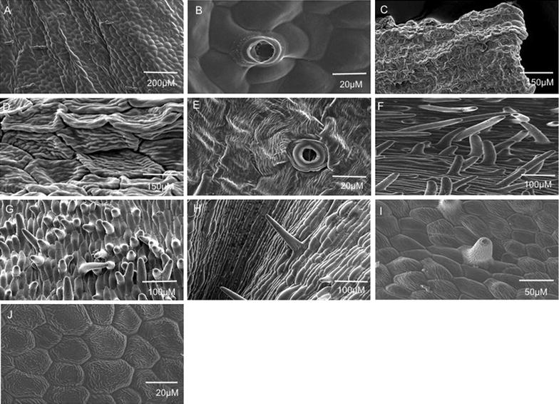
Fig. 2.

Surface morphology of Vanda Mimi Palmer floral parts. A, B stomata and trichomes on the sepal; C, D ridged adaxial epithelial cells on the labellum’s side-lobe; E stomata on the labellum; F, G dense protrusions/appendages that arise directly from the adaxial epidermal cells on the labellum’s mid-lobe; H long columnous adaxial epithelial cells on the labellum’s mid-lobe; I striated conical adaxial epithelial cells on the labellum’s mid-lobe; J polyhedral striated flattened adaxial epithelial cells
