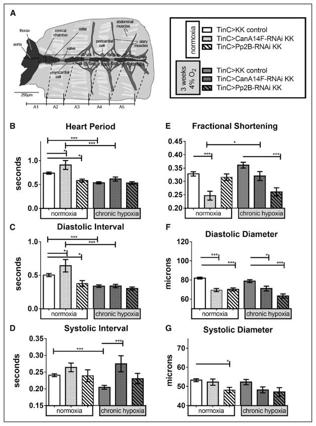
Figure 4.
Effect of chronic hypoxia on heart function in hearts with myocardial-specific knockdown of calcineurins. A, left, Diagram of Drosophila heart tube within dissected abdominal preparation (image courtesy of Dr. Georg Vogler). TinC-Gal4 drives expression specifically in myocardial cells (central tube), excluding the ostia (inflow valves). Hand4.2-Gal4-driven expression includes the ostia, as well as the pericardial cells. B–G, Heart function data under normoxia (left bars) or CH exposure (right bars) per genotypes indicated in figure key (A, right): progeny of CanA14F-RNAi and Pp2B-RNAi crossed to a TinC-Gal4 driver causes knockdown (KD) specifically in myocardial cells during embryonic development and in pupal stages through adulthood. Hearts were dissected and assayed under 21% O2 after 3 weeks at 21% or 4% O2 (as in Zarndt et al20). B, Heart period (HP) was elevated in CanA14F-RNAi but lowered in Pp2B-RNAi flies under normoxic conditions. After 3 weeks CH, HP was reduced for control and did not changed further on calcineurin KD (condition: F=14, P=0.0002; genotype: F=8.1, P=0.0004; interaction =2.7, P=0.070). C, Diastolic intervals changed similar to HP (condition: F=13, P=0.0004; genotype: F=3.8, P=0.025; interaction: F=2.1, P=0.13). D, Systolic intervals (SI) did not change on myocardial calcineurin KD under normoxia but were reduced in control flies after CH. Calcineurin KD in the myocardium under CH increased the systolic interval, similar to the Hand4.2-Gal4 data in Figure 3D (condition: F=0.64, P=0.43; genotype: F=7.8, P=0.0006; interaction: F=1.2, P=0.31). E, Fractional shortening (FS) was significantly reduced in myocardial CanA14F-RNAi KD, compared with controls, at normoxia and compared with CanA14F KD under CH. Myocardial Pp2B KD exhibited reduced FS on CH, but not under normoxia (condition: F=0.53, P=0.47; genotype: F=20, P<0.0001; interaction: F=9.0, P=0.0002). F, Diastolic diameters were decreased significantly with CanA14F-RNAi and Pp2B-RNAi KD under normoxia and CH (condition: F=7.3, P=0.0009; genotype: F=14, P=0.0003; interaction: F=7.3, P=0.0009). F, Systolic diameters were decreased, significantly only with Pp2B-RNAi KD at normoxia (condition: F=11, P=0.0009; genotype: F=1.4, P=0.26; interaction: F=3.0, P=0.055). All values are mean ± SEM (normoxia: N=32 KK control, N=30 CanA14F-RNAi, N=38 Pp2B-RNAi; chronic hypoxia: N=28 KK control, N=22 CanA14F-RNAi, N=20 Pp2B-RNAi). Data analysis: Kruskal–Wallis test and Dunn multiple comparisons post hoc test and 2-way analysis of variance (ANOVA) and Tukey’s multiple comparisons post hoc test; n.s.=not significant, *P<0.05, **P<0.01, ***P<0.001.

