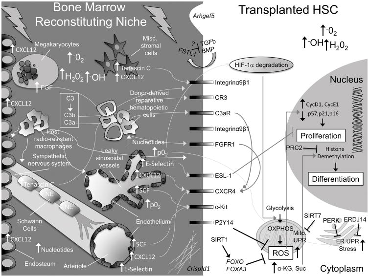
Figure 1. Summary of molecular alterations driven by classical pre-transplant conditioning regimens in HSCs and the bone marrow niche.
Here, we present a schematic to highlight some of the gross physical and molecular changes that occur in the bone marrow niche and within HSCs. For simplicity, not every known cellular component of the niche is pictured. In the niche, C3 is cleaved to C3b and C3a, which interact with HSC CR3 and C3aR receptors and stimulate homing by increasing, among other things, CXCR4. Megakaryocytes, which are attracted to the endosteum from sinusoidal vessels by increasing endosteal-CXCL12, also upregulate CXCR4 on HSCs via increased secretion of FGF. Schwann cells and stromal cells release TENASCIN C, which stimulates HSC migration and adhesion. Endothelial cells upregulate E-SELECTIN, CXCL12 and SCF. Sinusoidal vessels are damaged and leaky, resulting in an increase in O2 partial pressure (p02) and BM ROS levels. This contributes to H1F-1α degradation in HSCs, promoting their transition from glycolysis to oxidative respiration (OXPHOS), which further increases intracellular ROS levels. FOXOs, FOXA3, and signaling downstream of P2Y14 help HSCs cope with rising ROS levels. SIRT1 activates FOXOs. SIRT7 inhibits the increase in the mitochondrial unfolded protein response (UPR). Increased ROS stimulates HSC division and an ER-UPR. PERK and ERDJ14 counteract this effect in transplanted HSCs. Free nucleotide levels rise in the BM and are sensed by purinergic receptors, like P2Y14, which regulates ROS. Increasing intracellular α-Ketoglutarate (α-KG) promotes HSC differentiation via Histone demethylation. PRC2 complex counteracts this effect by promoting Histone methylation. Transplanted HSCs condition the reconstituting niche by secreting FSTL1 and extracellular matrix components (via Crispld1) and (very likely) additional factors (e.g. IL-8). Transplanted hematopoietic cells facilitate recovery of the conditioned niche. Figure Key: the bone marrow space is depicted on a dark gray background, the HSC intracellular space is light gray, and the HSC nucleus is dark gray. Major cell types are labeled in white font, major changes in the bone marrow space are labeled in white font, and major changes in the HSC are labeled in black font.

