Figure 1.

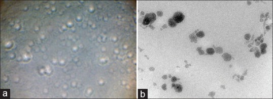
(a) Phase contrast microscopy image of transferosomal formulation (S7), (b) TEM image of transferosomal formulation (S7) (reproduction size at column width)

(a) Phase contrast microscopy image of transferosomal formulation (S7), (b) TEM image of transferosomal formulation (S7) (reproduction size at column width)