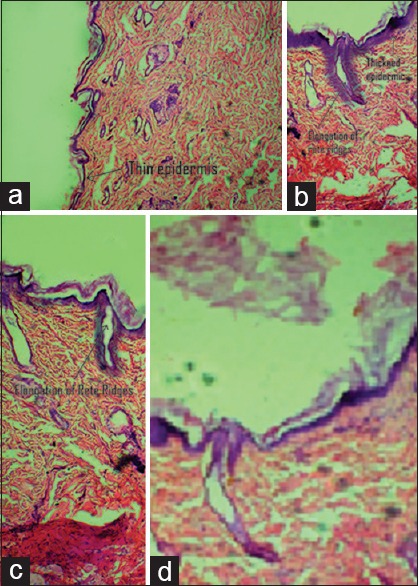
Figure 2.

(a) Histopathology image of control animal's skin section with thin epidermis, (b) histopathology image of disease-induced (imiquimod treated) skin showing thickened epidermis, elongation of rete ridges, (c) histopathology image of animal treated with conventional gel showing nominal reduction in epidermis and rete ridges, (d) animal treated with Berberis aristata extract loaded transferosomal gel marked reduction in epidermal thickness and elongation of rete ridges (reproduction size: all at column width)
