
Figure 1.
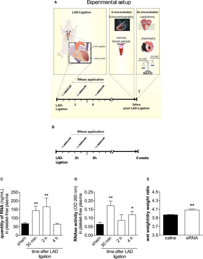
eRNA and intrinsic RNase activity in mice after the induction of myocardial infarction. A and B, A diagram of the study protocol of short‐term (A) and long‐term (B) assessments. C, Quantification of eRNA in platelet‐free plasma samples of mice 24 hours after ligation of the LAD (**P<0.01 vs sham‐operated mice; n=7). D, RNase activity in platelet‐free plasma following ligation of the LAD (*P<0.05, **P<0.01 vs sham‐operated mice; n=7). E, Wet/dry ratios of mouse hearts following intravenous application of external eRNA (15 μg/kg) or saline (*P<0.05, n=7). eRNA indicates extracellular RNA; LAD, left anterior descending coronary artery; OD, optical density; RNase, ribonuclease.