
Figure 6.
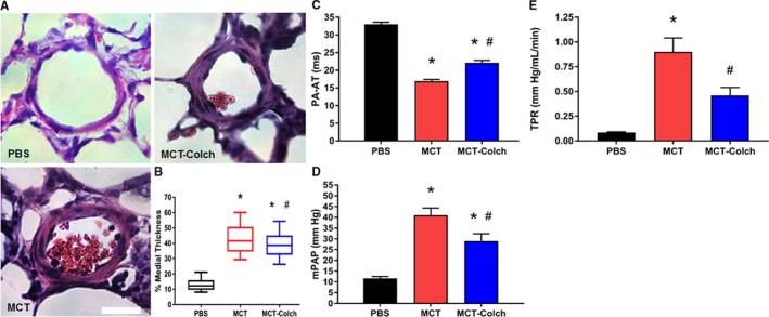
Colchicine reduces severity of pulmonary vascular disease in MCT rats. A, Representative hematoxylin and eosin–stained lung sections showing pulmonary arterioles. B, Box‐and‐whisker plots of percentage medial thickness (PBS 13.5±0.6% n=85 arterioles from 3 different animals, MCT 43.4±1.5% n=78 arterioles from 3 animals, MCT‐colchicine 39.5±1.1% n=81 arterioles from 3 different animals). Whiskers extend from 10th to 90th percentiles. Colchicine reduces pulmonary arteriolar medial thickness. C, Colchicine increases PA acceleration time (PBS 33±1 milliseconds, n=16, MCT 17±1 milliseconds, n=21, MCT‐colchicine 22±1 milliseconds n=22). Quantification of (D) mPAP (PBS 12±1 mm Hg, n=5, MCT 41±3 mm Hg, n=13, MCT‐colchicine 29±3 mm Hg n=14) and (E) TPR (PBS 0.1±0.01 mm Hg/[mL·min], n=5, MCT 0.9±0.1 mm Hg/[mL·min], n=9, MCT‐colchicine 0.5±0.1 mm Hg/[mL·min], n=11) from right heart catheterization. Colchicine improves pulmonary hemodynamics. *Significantly different from PBS, #significantly different from MCT rats as determined by 1‐way ANOVA with Tukey post hoc analysis. Scale bar 25 μm. Colch indicates colchicine; MCT, monocrotaline; mPAP, mean pulmonary arterial pressure; PBS, phosphate‐buffered saline; TPR, total pulmonary resistance.