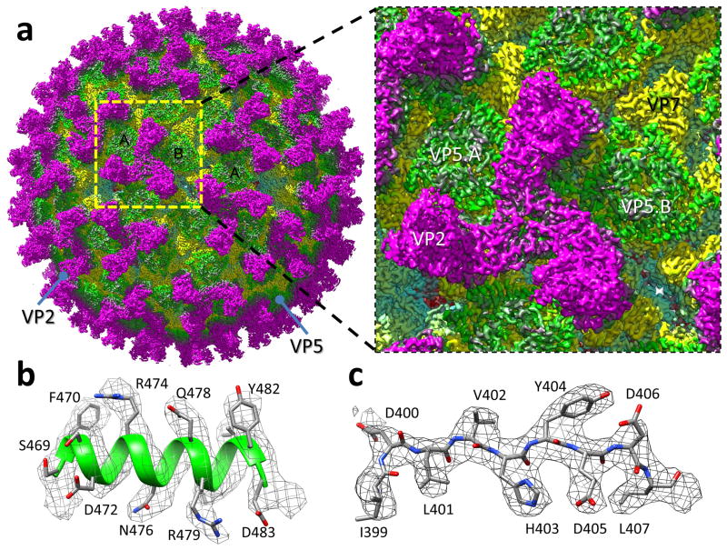
Figure 1. CryoEM reconstruction of the BTV virion.
(a) CryoEM density map of the BTV virion shown as radially-coloured surface representation, and a close-up view of the boxed area containing an asymmetric unit. (b,c) Close-up views of the α17 helix (b) and a β-strand of VP5 (c), showing side-chain density of amino-acid residues in a helix and a loop, respectively. The atomic model is shown as ribbons or sticks superimposed with the density (mesh).

