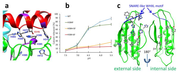
Figure 5. Membrane interaction elements of VP5.
(a) The histidine cluster of VP5. The fully conserved Glu316 is also highlighted. (b) Membrane permeability as measured by calcein release from liposomes as a function of pH for WT, and single- (H384F), double- (H384-5F) and triple- (H384-6F) mutants of the histidine His384–386 cluster of VP5. Error bars represent s.d. of n = 3 technical repeats. (c) External (viewed along the arrow in Figure 3b and internal sides of the core β-meander motif of the anchoring domain, showing a cluster of 5 and 7 aromatic residues on the internal and external side respectively. The SNARE-like WHXL motif (W411-L414) located on the β7 strand is shown in blue.

