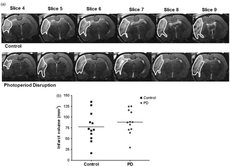
Figure 5.
T2-weighted MRI images taken at 48 h post MCAO showing hyper-intense (highlighted) region as infarcted area. Representative slices from median animal demonstrate comparable ischemic damage in both groups (a). Photoperiod disruption did not increase infarct volume in PD rats (b). Data points indicate individual rats and the horizontal bar represents the mean (p > 0.05, Student unpaired t test). PD: photoperiod disruption.

