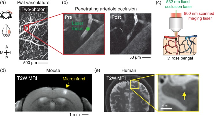
Figure 1.
Modeling of microinfarcts in mice by optical occlusion of single cortical penetrating arterioles in vivo. (a) Wide-field two-photon imaging of the pial vasculature through a thinned-skull cranial window. A = anterior, L = lateral, P = posterior, M = medial. (b) High-resolution imaging and focal photothrombotic occlusion of a single penetrating arteriole. Green circle shows location of focused 532 nm laser irradiation. (c) Cartoon describing focal 532 nm laser activation of circulating Rose Bengal dye in targeted arterioles. Imaging is performed with an 800 nm scanned Ti-sapphire laser. (d) Coronal view of microinfarct resulting from occlusion of a single penetrating arteriole, viewed with T2-weighted 7 T MRI 24 h post-occlusion. (e) A cortical microinfarct identified in the living human brain using T2-weighted 7 T MRI. Data reproduced with permission from van Veluw et al.33

