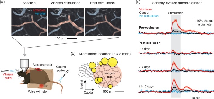
Figure 6.
Arteriole reactivity following strategic placement of microinfarcts in barrel cortex. (a) Awake, head-fixed mice were imaged with two-photon microscopy to examine pial and penetrating arteriole dilation in response to whisker stimulation, i.e. functional hyperemia. As a control for general arousal, air puffs were delivered to the tail. (b) In each mouse (n = 8), a single microinfarct was strategically placed to flank the primary barrel cortex. The positions of all eight microinfarcts (yellow) are shown as a composite. (c) Sensory-evoked arteriole dilation pre-occlusion and at various periods following penetrating arteriole occlusion. N = 36 arterioles (15 surface arterioles, 21 penetrating arterioles) over eight mice. Data are mean ± SEM.

