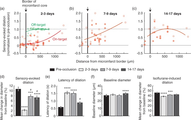
Figure 7.
Disruption of single-vessel hemodynamics in peri-lesional tissues surrounding microinfarcts. (a–c) Sensory-evoked dilation as a function of distance from the microinfarct core (red; n = 50, 36, and 24 arterioles over six mice for panels a, b, and c, respectively). Open circles represent surface arterioles, and filled circles represent penetrating arterioles. An off-target control group showed no change in dilatory function beyond the region of photothrombotic irradiation (green in panel a; n = 33 arterioles over three mice). Lines correspond to second order polynomial regression fits of the data. (d) Mean change in diameter during whisker stimulation over a group of arterioles that could be measured repeatedly over all time-points (p < 0.0001 main effect, Friedman statistic = 82.1, Friedman test with repeated measures; Dunn’s post hoc yields ****p < 0.0001 or ***p < 0.001, compared to pre-occlusion, and #p < 0.05, compared to 2 to 3 days; n = 36 arterioles over six mice). Data are mean ± SEM. (e) Latency to peak dilation after onset of stimulation (p < 0.0001 main effect, Friedman statistic = 82.6, Friedman test with repeated measures; Dunn’s post hoc yields ****p < 0.0001, compared to pre-occlusion, and #p < 0.05, compared to 2 to 3 days and 7 to 9 days). (f) Baseline diameter of arterioles prior to stimulation. (p < 0.0001 main effect, F(2.42,84.58) = 8.96, one-way ANOVA with repeated measures; Tukey’s post hoc yields ****p < 0.0001, compared to pre-occlusion). (g) Mean change in diameter after inhalation of vasodilating anesthetic isoflurane (3% MAC in air) (p < 0.0001 main effect, Friedman statistic = 39.9, Friedman test with repeated measures; Dunn’s post hoc yields ****p < 0.0001 or ***p < 0.001, compared to pre-occlusion).

