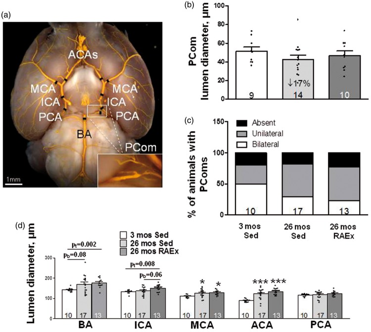
Figure 2.
Lumen diameters of primary intracranial arteries increase with aging and exercise-training further augments this. (a) Black hashes identify site of measurement (250 µm from bifurcations; at midpoint for ICA, PCA and PComs). Statistics symbols are defined in the legend for Figure 1; the values above the bracketed two-group comparisons for BA and ICA are double the Bonferroni value, computed for the comparisons defined in Figure 1 legend, to protect against a type-II error in the two-group comparison. (b, c) Diameter and number of PComs (% of animals with PComs present bilaterally, unilaterally or absent) did not change with aging or exercise-training (ANOVA for diameter p = 0.20, two-tailed X2 test for percentage of animals with absent + unilateral p = 0.25). Panel D, ANOVAs for BA, ICA and MCA are p < 0.05, ACA p < 0.001. pb = p-value for Bonferroni constrained to two comparisons (26 mos Sed or 26 mos RAEx vs. 3 mos Sed). pt = p-value for one-tailed t-test.

