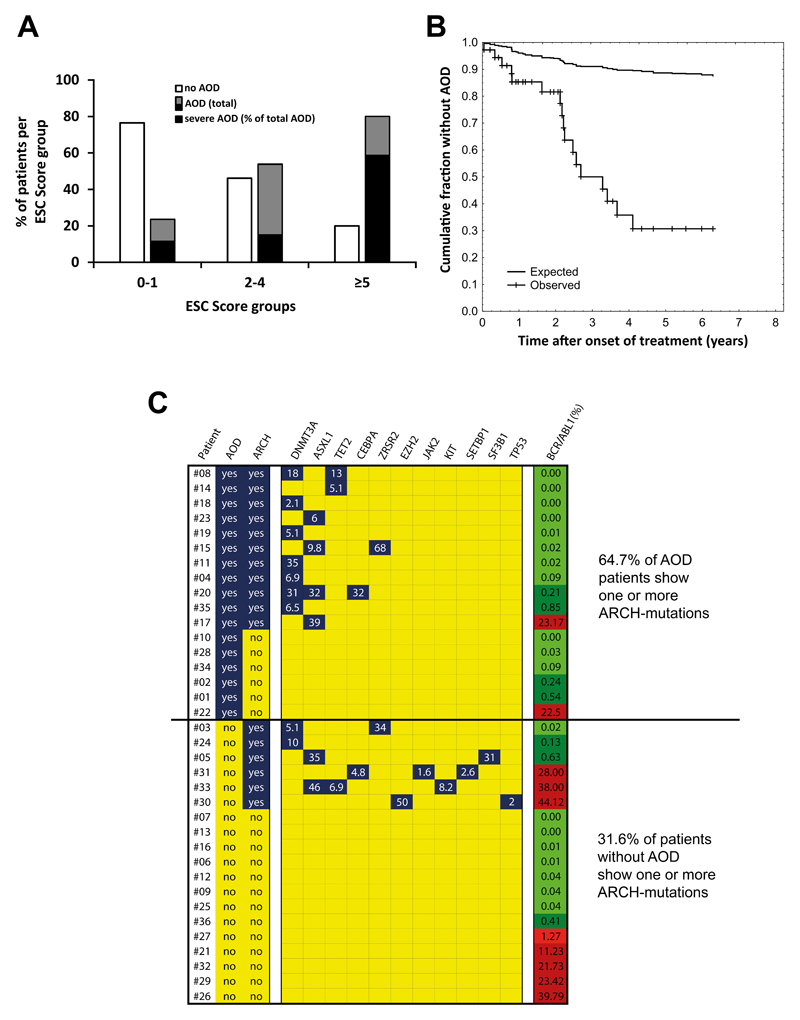
Figure 4. Risk factors for vascular events in TKI-treated patients.
(A) Our Nilotinib-treated patients were separated into three risk-groups according to the European Society of Cardiology (ESC) SCORE. The numbers of patients in each ESC-cohort were: 17 in ESC 0-1; 13 in ESC 2-4; and 5 in ESC ≥5. The percentage of patients without AOD (open bars) and with AOD (black and grey bars) is shown for each group. The black parts of the black and grey bars represent the percentages of severe AOD events relative to total AOD events. (B) The curves show the reduction of the cumulative fraction of individuals without AOD in our Nilotinib-treated patients and a risk-factor-matched population in high-income countries described in a meta-analysis by Fowkes et al.39. The difference in reduction of the cumulative fraction between the two groups is statistically significant by log-rank test: p<0.05. (C) Overview of ARCH-related gene variants in all 36 Nilotinib-treated patients. Peripheral blood samples were analyzed for the presence of ARCH mutations at the time of best response. White numbers indicate the variant allele frequencies. The right column shows BCR-ABL1 mRNA levels (% of ABL1) at the time of sampling; levels lower than 1% are depicted in green, levels above 1% in red. The difference in the percentages of patients carrying ARCH mutations between the two groups (AOD patients versus patients without AOD) was significant (p<0.05) as assessed by the chi-squared test. AOD: Arterial occlusive disease; ARCH: age-related clonal hematopoiesis (at least one ARCH-mutation detected).

