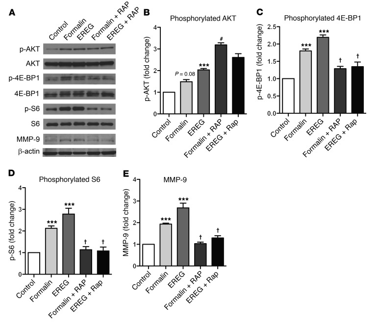
Figure 6. EREG and formalin induce phosphorylation of AKT and 4EBP-1 and increase the expression of MMP-9 in lumbar DRG tissue.
EREG (10 ng, i.t.) or 5% formalin (20 μl, intraplantar) was injected and lumbar DRG tissue harvested 40 minutes later. Rapamycin was injected 20 minutes before EREG or formalin to mimic behavioral experiment parameters. (A) Representative Western blots showing the phosphorylated and total protein abundance for AKT, 4E-BP1, and S6. The total amount of MMP-9 is also presented. Quantification (phosphorylated/total) for the percentage of fold increase (compared with the control condition) in phosphorylated AKT, 4E‑BP1, and S6 is presented in panels B–D along with the quantification for total MMP-9 (E). Bars represent mean ± SEM for relative change in protein expression. (B) EREG significantly increases the phosphorylation of AKT in DRG tissue. (C) Both formalin and EREG increase the phosphorylation of 4E-BP-1, and the increases are blocked by rapamycin. (D) The phosphorylation of S6 is significantly elevated relative to control tissue by EREG treatment, an increase blocked by rapamycin. (E) Formalin and EREG significantly increase MMP-9 expression, and these increases are blocked by rapamycin. Sample sizes in all groups are n = 4–6. One-way ANOVA for all panels followed by Dunnett’s case-comparison post hoc test. ***P < 0.001 compared with control tissue. †P < 0.05 decrease compared with EREG or formalin alone group. #P < 0.05 increase compared with EREG or formalin alone group.

