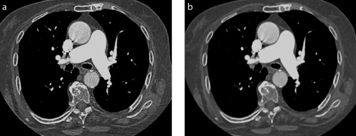
Figure 1. a, b.
Axial computed tomography (CT) images reconstructed with filtered back projection (FBP) (a) or iterative reconstruction (IR) (b) in an oncologic patient with mild dyspnea who underwent CT pulmonary angiography to exclude pulmonary embolism. Axial image reconstructed with IR is waxier; however, the information is essentially the same, excluding pulmonary embolism in the main pulmonary arteries.

