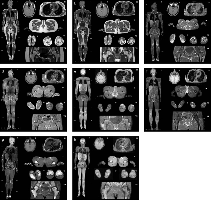
Figure 1. a–h.
Magnetic resonance images of patients with generalized lipodystrophy. Panels (a, b) show control subjects with normal adipose tissue distribution (a, a 28-year-old healthy woman; b, a 26-year-old healthy man). Panels (c, d) show adipose tissue distribution in patients with CGL1: (c), a 30-year-old female (patient 1.1); (d), a 31-year-old male (patient 2.2). Panels (e, f) show adipose tissue distribution in patients with CGL 2: (e), a 25-year-old female (patient 6.1); (f), a 19-year-old male (patient 6.2). Panel (g) shows adipose tissue distribution in a 16-year-old female patient with CGL4 (patient 8.1). Panel (h) shows adipose tissue distribution in an 11-year-old female patient with AGL (patient 10).
(I), whole body, coronal T1-weighted imaging; (II), orbital fat and scalp, axial T1-weighted imaging; (III), breasts, axial T1-weighted imaging; (IV), external genital region and palms, axial T1-weighted imaging; (V), soles, axial T1-weighted imaging; (VI), patellar region, axial T1-weighted imaging; (VII), hip region, coronal T1-weighted imaging.

