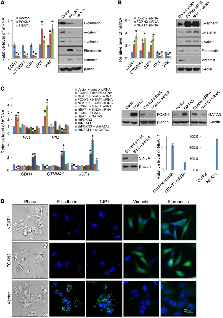
Figure 5. The FOXN3-NEAT1-SIN3A complex promotes EMT.
(A) MCF-7 cells were transfected with FOXN3 or NEAT1 for the measurement of the indicated epithelial/mesenchymal markers by qPCR or Western blotting. (B) MCF-7 cells were transfected with FOXN3 siRNA or NEAT1 siRNA for the measurement of the indicated epithelial/mesenchymal markers by qPCR or Western blotting. (C) MCF-7 cells were transfected with the indicated siRNAs and/or expression vectors for the measurement of the indicated epithelial/mesenchymal markers by qPCR. The efficiency of knockdown or overexpression was verified by Western blotting or qPCR. In A–C, error bars represent mean ± SD for triplicate experiments (*P < 0.05, 2-way ANOVA). (D) MCF-7 cells were transfected with FOXN3 or NEAT1 for the morphological examination by phase-contrast microscopy. The epithelial (E-cadherin and TJP1) and mesenchymal (vimentin and fibronectin) markers were immunofluorescently stained and analyzed by confocal microscopy. DAPI staining was included to visualize the nucleus (blue). Representative images from triplicate experiments are shown.

