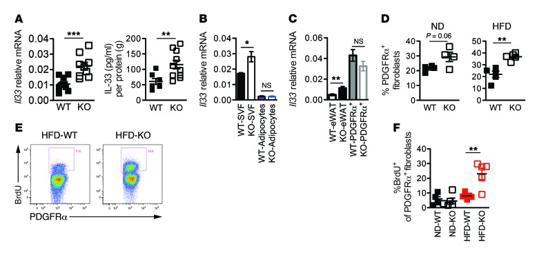
Figure 4. PDGFRα+ fibroblast expansion accounts for higher expression of IL-33 in cad-11–/– adipose tissue.
(A) IL-33 expression in adipose tissue of WT and cad-11–/– mice fed a HFD for 5 weeks (left graph: n = 9 HFD-WT and n = 9 HFD-KO, pooled from 2 independent experiments; right graph: n = 7 HFD-WT and n = 10 HFD-KO, pooled from 2 independent experiments). (B) Il33 mRNA expression in SVF cells and adipocytes isolated from adipose tissue of mice fed a HFD for 12 weeks (n = 4 WT-SVF, n = 6 KO-SVF, n = 5 WT-adipocytes, and n = 7 KO-adipocytes, pooled from 2 independent experiments). (C) Il33 mRNA expression in adipose tissue and flow-isolated PDGFRα+ fibroblasts (n = 5 WT-eWAT, n = 5 KO-eWAT, n = 5 WT-PDGFRα+, and n = 5 KO-PDGFRα+). Data are representative of 2 independent experiments. (D) Percentage of PDGFRα+ fibroblasts in SVF cells from mice fed a ND or HFD for 5 weeks (n = 4 ND-WT, n = 4 ND-KO, n = 5 HFD-WT, and n = 5 HFD-KO). Results are representative of more than 3 independent experiments. (E) Representative flow cytometric plots of BrdU uptake in PDGFRα+ fibroblasts from WT and cad-11–/– mice fed a HFD for 1 week. (F) Percentage of BrdU+ cells among PDGFRα+ fibroblasts (n = 5 ND-WT, n = 5 ND-KO). Results are representative of 2 independent experiments. Data are expressed as the mean ± SEM. *P < 0.05, **P < 0.01, and ***P < 0.001, by unpaired Student’s t test (A–D and F).

