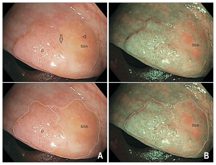
Fig. 4.
Endoscopic appearance of sessile serrated adenomas/polyps (SSA/Ps) with dysplasia. A 20 mm SSA/P-D viewed under white light (A) and narrow band imaging (B) with and without the dysplastic (label D) and nondysplastic (label SSA) components outlined. The lesion has developed a raised, nodular component on the left-hand aspect with a type IV surface pit pattern indicative of dysplastic transformation (label D). The nondysplastic component of the lesion (label SSA) is pale with relatively hypovascular background surface markings and is covered by a thin layer of stool debris (arrowhead). Note there is an obvious transition zone from the nondysplastic flat SSA/P to the area of dysplasia (arrow). The lesion and a rim of normal tissue were removed en bloc by endoscopic mucosal resection; histology confirmed a completely resected SSA/P with mild dysplasia.

