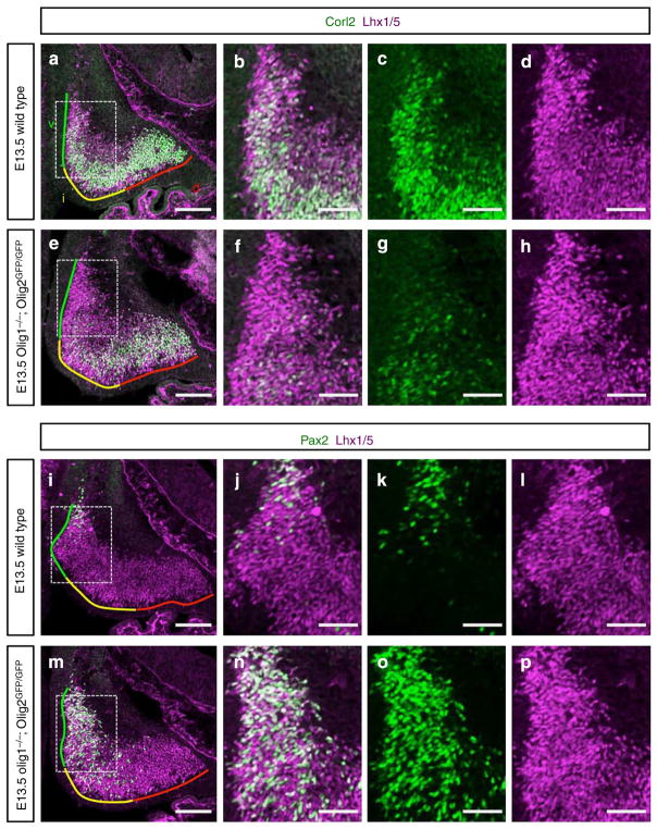
Figure 5. Phenotypes of Olig1/2 dKO mice.
(a,e) Cerebellar primordium of E13.5 (a) wild-type and (e) Olig1/2 dKO mouse stained with antibodies for Corl2 and Lhx1/5. Coloured lines indicate ventral (green), intermediate (yellow) and dorsal (red) regions of cerebellar primordia, respectively. (b–d,f–h) Higher-magnification images of the rectangular regions in (a) and (e), respectively. (i,m) Cerebellar primordium of E13.5 (i) wild-type and (m) Olig1/2 dKO mouse stained with antibodies for Pax2 and Lhx1/5. (j–l,n–p) Higher-magnification images of the rectangular regions in (i) and (m), respectively. Scale bars represent (a,e,i and m) 100 μm, (b–d,f–h,j–l and n–p) 50 μm.

