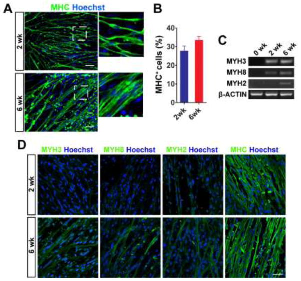
Figure 3. Longer differentiation supports more mature phenotype in myotubes.
(A) Representative image of MHC+ myotubes. From 2 to 6 weeks, single nucleated nascent myotubes migrated and fused to form larger multinucleated MHC+ myotubes. Scale bar = 50 μm. (B) The number of MHC+ cells tended to be increased at 6 weeks, although the difference did not reach statistically significance. (C) RT-PCR analysis of MYH genes in differentiated cells. Specific primers were used to detect the gene expression of embryonic (MYH3), fetal/perinatal (MYH8), and adult form of MHC (MYH2). (D) Immunocytochemistry of MYH3, MYH8, MYH2, and MHC (with MF20 antibody clone) in differentiated myotubes. Scale bar = 50 μm.

