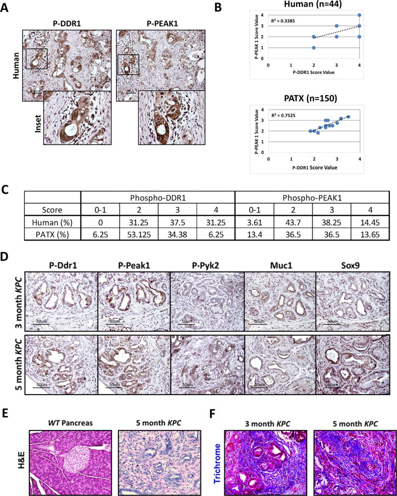
Figure 1. DDR1 signaling in human and mouse PDA.
(A) Immunohistochemical detection of phospho-DDR1 and phospho-PEAK1 in human PDA. TMAs of primary human PDA (44 samples) demonstrated phospho-DDR1 and phospho-PEAK1 localized to similar regions. (B) Pearson correlation of p-PEAK1 and p-DDR1 expression in human and patient-derived tumor xenograft (PATX) TMA samples. (C) Percent of TMA samples positive for p-DDR1 and p-PEAK1. Scoring system is denoted as: low or no reactivity (0–1), moderate reactivity (2), strong reactivity (3), and very strong reactivity (4). (D–F) Histological analyses of the KPC (LSL-KrasG12D/+; LSL-Trp53R172H/+; p48Cre/+) GEMM of PDA. (D) Immunohistochemical detection of phospho-Ddr1, phospho-Peak1, phospho-Pyk2, Muc1, and Sox9 in KPC tumors. Ddr1 activation and downstream signaling through effectors such as Peak1 and Pyk2 was present in early PanIN lesions (similar to regions positive for Muc-1 staining) and in advanced adenocarcinoma (similar to regions of Sox9 staining). Tissue from an early (3-month) and advanced (5-month) stage of the KPC model was evaluated. (E) H&E histology of normal WT pancreas and PDA in a 5-month-old KPC mouse. (F) Trichrome analysis of PDA from 3- and 5-month-old KPC animals.

