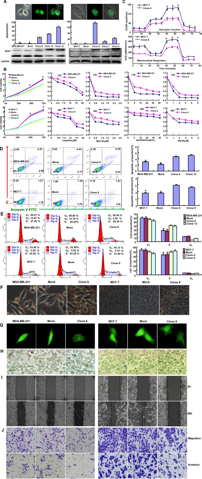
Figure 1. ING5 expression altered the phenotypes of breast cancer cells.
After transfection of pCDNA3.1-ING5, its expression became strong in MDA-MB-231 and MCF-7 cells by fluorescence, RT-PCR and Western blot (A). The cell viability was measured using MTT assay in both breast cancer cells and their ING5 transfectants, even treated by cisplatin, MG132, paclitaxel and SAHA (B). The glucose metabolism of MCF-7 and its transfectant was detected by XF-24 extracellular flux analyzers (C). The apoptosis, cell cycle, fat accumulation, autophagy, senescence, migration and invasion were examined by Annexin-V staining (D), PI staining (E), Oil red O staining (F), the transient transfection of LC-3B-expressing plasmid (G), β-galactosidase staining (H), wound healing (I), and transwell chamber assay (J) *p < 0.05, compared with the transfectants.

