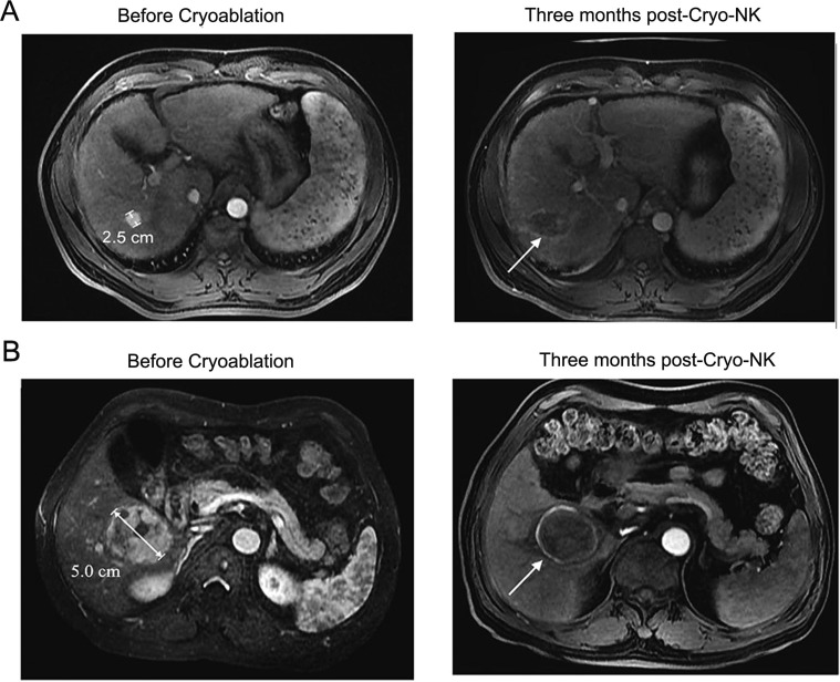
Figure 4. MRI images of 2 representative cases achieved CR at three months post-Cryo-NK.
(A) Case #1, a 50-year-old male, stage III, a maximum HCC nodule of 2.5 cm, MRI showed no enhancement in the occupying lesion, with mild shrinkage of the area; (B) Case #2, a 48-year-old female, stage IV, a maximum HCC nodule of 5.0 cm, MRI showed a lesion with a large area of necrosis.

