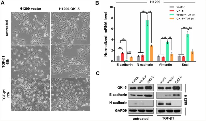
Figure 3. QKI-5 overexpression restrains the TGF-β1-induced EMT of LC cells.
(A) Representative images of H1299-vector (control) and H1299-QKI-5 (QKI-5-overexpressing) cells untreated or incubated with TGF-β1 (5 ng/mL) for 48 or 72 h (×200). (B) mRNA levels of E-cadherin, N-cadherin, Vimentin and Snail in the indicated cells incubated with TGF-β1 or untreated in DMEM for 72 h as in A. The mRNAs were detected by qRT-PCR and normalized against GAPDH. The ratios of the above mRNAs to GAPDH mRNA in control cells (vector) were arbitrarily set to 1.0. The data are presented as the mean ± SD. **P<0.01, ***P<0.001. (C) Protein levels of QKI-5, E-cadherin and N-cadherin measured by Western blotting in the indicated H1299 cells.

