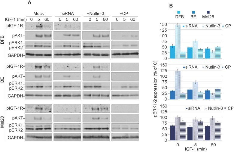
Figure 3. Characterization of balanced versus biased targeting strategies on IGF-1R signalling.
(A) DFB, BE and Mel28 melanoma cells were either transfected with siRNA towards IGF-1R (48 h) or treated with 1 μM Nutlin-3 (24 h) or 100 ng/mL CP (24 h), alongside mock controls. Serum starved cells were stimulated with IGF-1 (50 ng/mL) for 0, 5 and 60 min. Cell lysates were analyzed by WB for phosphorylated (p) versions of IGF-1R, Akt and ERK1/2, alongside GAPDH as a loading control. (B) pERK1/2 signals were quantified by densitometry, normalized to total ERK1/2 and expressed as a % of pERK1/2 in the mock-treated cells for each time point. Data correspond to the mean ± S.E.M. from three independent experiments.

