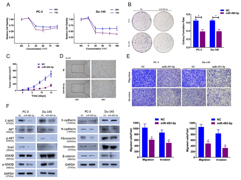
Figure 2. Effect of miR-493-5p on prostate cancer cell proliferation and cell motility.
(A) CCK-8 assay. The relative cell viability of the miR-493-5p-treated groups of PC-3 and Du-145 cells was lower than that of NC-treated groups (cell viability of 0 nM was regarded as 1.0). (B) Colony-formation assay (representative wells are presented). The colony-formation rate was lower for miR-493-5p (50 nM)-transfected groups compared to NC (50 nM)-transfected groups. (C, D) Tumor xenograft model. Tumor growth curves indicated that tumors in the miR-493-5p group grew more slowly. Decreased Ki-67 expression was also detected in miR-493-5p-treated tumors. (E) Transwell assay (representative micrographs are presented). miR-493-5p (50 nM) impaired the motility of PC-3 and Du-145 cells. (F) Western blot analysis. miR-493-5p (50 nM) inhibited EMT and AKT/GSK-3β signaling-related protein expression in PC-3 and Du-145 cells. Error bars represent the S.E. obtained from three independent experiments; *P<0.05. Scale bar = 100 μm.

