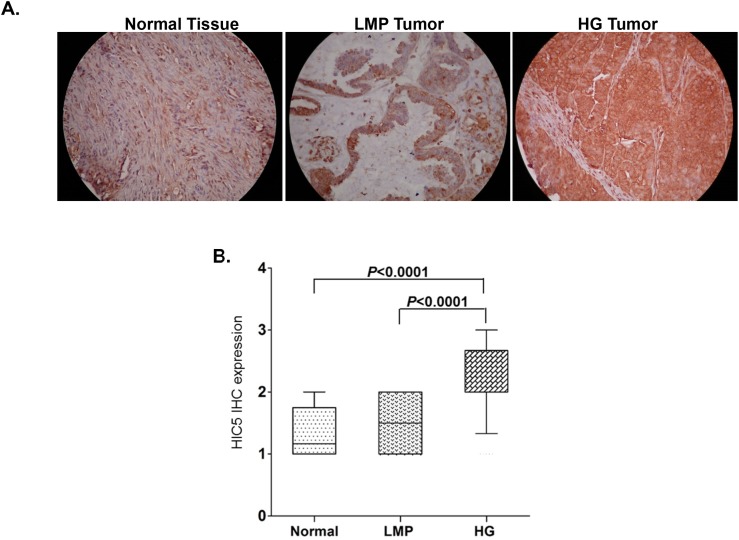
Figure 1. Analysis of Hic-5 expression in serous EOC tumors by IHC.
(A) Representative IHC images of Hic-5 protein expression in normal ovarian tissues, low-malignant potential (LMP) tumors and high-grade (HG) tumors. (B) Box-plot presentation of Hic-5 protein expression levels in normal ovarian tissues, low-malignant potential (LMP) tumors and high-grade (HG) tumors.

