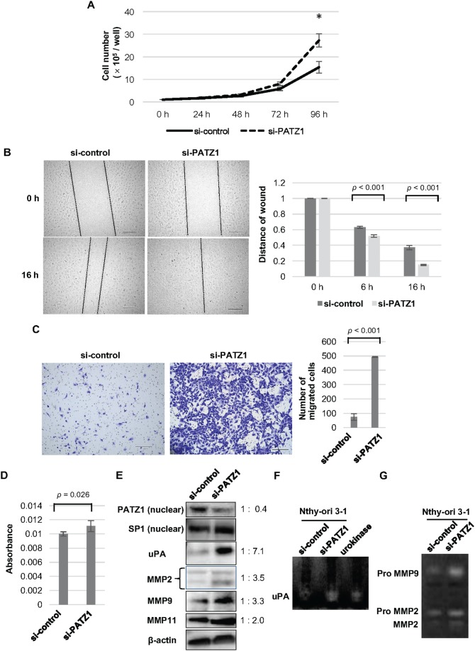
Figure 4. Effects of PATZ1 knockdown on cell proliferation, migration, and invasion in Nthy-ori 3-1 cells.
(A) Cell proliferation assay for Nthy-ori 3-1 cells transfected with control siRNA (si-control) or siRNA targeting PATZ1 (si-PATZ1). The number of cells was counted every 24 h until 96 h after seeding the cells. The number of cells is shown in the bar chart. *p < 0.05. (B) Scratch wound assay for Nthy-ori 3-1 cells transfected with si-control or si-PATZ1. Representative images of scratch wound assay in Nthy-ori 3-1 cells transfected with si-control or si-PATZ1 at 0 h and 16 h after a confluent cell monolayer was scratched (left panel, scale bar = 200 μm). The length of the gap at 100 points was measured for each sample and the average ratio of residual gap to the initial gap is shown in the bar chart (right panel). (C) Chamber migration assay for Nthy-ori 3-1 cells transfected with si-control or si-PATZ1. Representative images of migrating cells stained with crystal violet in Nthy-ori 3-1 cells transfected with si-control or si-PATZ1 at 24 h (left panel, scale bar = 200 μm). The number of migrated cells is shown in the bar chart (right panel). (D) Chamber-invasion assay for Nthy-ori 3-1 cells transfected with si-control or si-PATZ1. The cells that invaded through the transwell chamber coated with collagen type IV were counted as described in Materials and Methods. The absorbance values are shown in the bar chart. (E) The expression of PATZ1, uPA, and MMPs in Nthy-ori 3-1 cells transfected with si-control or si-PATZ1. A representative western blot is shown. SP1 and β-actin were used as internal loading control for nuclear extract and whole cell lysate, respectively. (F) Activity of uPA in Nthy-ori 3-1 cells transfected with si-control or si-PATZ1. A representative picture of fibrin zymography is shown. Urokinase was used as a positive control and the enzymatic activity was detected at 42 kDa. (G) Activities of MMP2 and MMP9 in Nthy-ori 3-1 cells transfected with si-control or si-PATZ1. A representative picture of gelatin zymography is shown. Degradation of gelatin is detected at approximately 92 kDa (Pro MMP9), 72 kDa (ProMMP2), and 66 kDa (MMP2).

