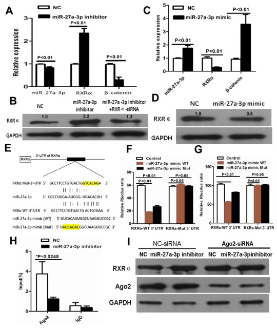
Figure 5.

(A) miR-27a-3p inhibitor increased RXRα mRNA and suppressed β-catenin mRNA expression in HCT116 by real-time PCR. (B) miR-27a-3p inhibitor increased RXRα protein expression in HCT116, however, RXRα knockdown reversed elevated RXRα protein expression which was induced by miR-27a-3p inhibitor in HCT116 by western blot analysis. The relative quantification of bands in Western blots was a ratio neutralized to GAPDH. (C) miR-27a-3p mimic suppressed RXRα mRNA and increased β-catenin mRNA expression in SW480 by real-time PCR. (D) miR-27a-3p mimic suppressed RXRα protein expression in SW480 by western blot analysis. The relative quantification of bands in Western blots was a ratio neutralized to GAPDH. (E) Predicted binding sites of miR-27a-3p in the wild type 3’-UTR of RXRα. Mutations in the 3’-UTR of RXRα and miR-27a-3p mimic were highlighted in yellow. (F-G) miR-27a-3p significantly inhibited the luciferase activities of RXRα-WT 3’UTR reporter in HCT116 (F) and SW480 (G) cells. However, miR-27a-3p had no effect on the luciferase activities of RXRα-Mut 3’UTR reporter in HCT116 (F) and SW480 (G) cells. (H) RNA binding protein immunoprecipitation assay showed Ago2 was associated with miR-27a-3p. (I) Ago2 knockdown dramatically decreased the effect of miR-27a-3p on RXRα expression in HCT116 cells.
