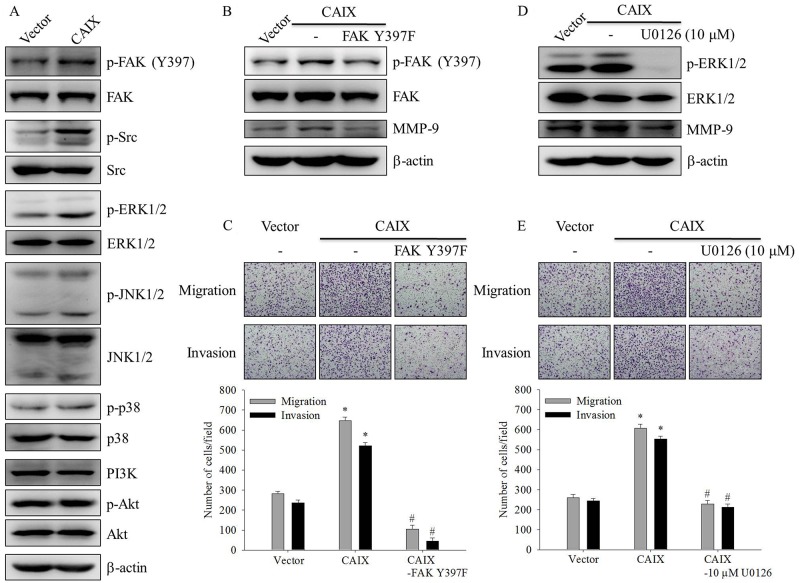
Figure 4. The FAK/Src and ERK signaling pathways are crucial for CAIX-induced MMP-9 upregulation and cell migration and invasion.
(A) The levels of total and phosphorylated FAK, steroid receptor coactivator (Src), ERK1/2, JNK1/2, p38, and Akt in SCC-9 cells with or without CAIX expression were determined using Western blot analyses of whole cell lysates. b-actin was used as a loading control. (B) SCC-9 cells with CAIX expression were treated with FAK Y397F for 24 h. Whole cell lysates were collected, and the expressions of FAK and MMP-9 were determined using Western blotting. b-actin was used as a loading control. (C) Cells treated with FAK Y397F for 24 h were analyzed using the Boyden chamber assay. (upper) Images were obtained using an inverted contrast light microscope under 100× magnification. (lower) The number of migrated and invaded cells per well was counted in three arbitrary visual fields. The results are expressed as mean ± SD of three replications. *p < 0.05 versus vector control; #p < 0.05 versus CAIX expression. (D) SCC-9 cells with CAIX expression were treated with 10 mM U0126 for 24 h. Western blotting was used to examine the effects of U0126 on ERK and MMP-9 expression. b-actin was used as a loading control. (E) Cells treated with 10 mM U0126 for 24 h were seeded into the Boyden chamber and were incubated at 37°C for 16 h. (upper) Images were obtained using an inverted contrast light microscope under 100× magnification. (lower) The number of migrated and invaded cells per well was counted in three arbitrary visual fields. The results are expressed as mean ± SD of three replications. *p < 0.05 versus vector control; #p < 0.05 versus CAIX expression.

