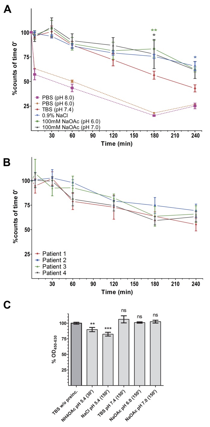
Figure 3. Radio- and protein-stability of 99mTc-DTPA-can225IgG.
(A) % of intact 99mTc-DTPA-can225IgG in various buffers, normalized to time point 0. Incubation was carried out at room temperature. Dotted pink line: PBS (pH 8.0) (n=2), Dotted orange line: PBS (pH 6.0) (n=2), Solid red line – TBS (pH 7.4), blue – 0.9% NaCl, green – NaOAc pH 6.0, grey – NaOAc pH 7.0. Denoted significances refer to the buffer indicated by the color, compared to TBS; n=3. (B) % of intact 99mTc-DTPA-can225IgG in canine mammary carcinoma patient’s sera, samples were incubated at 37°C; n=4. (C) % Captured can225IgG from of pre-incubated buffer samples, normalized to freshly prepared can225IgG. Statistical significances refer to differences in bound can225IgG compared to the reference sample; n=3.

