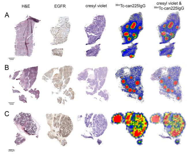
Figure 4. Immunohistochemistry and autoradiography of canine mammary carcinoma sections.
5 μm tumor sections of 3 canine mammary carcinoma patients (A-C) were stained with hematoxylin/eosin (first column) and for EGFR expression (second column). 10 μm sections of the same tumor were used for autoradiography with 99mTc-DTPA-can225IgG (fourth column). The same sections used for autoradiography were stained with cresyl violet in order to visualize tissue morphology (third column) and were used to generate an overlay (fifth column).

