
Figure 1.
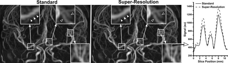
Comparison of ungated 3-shot radial QISS MRA obtained with standard and super-resolution reconstruction in a 34-year-old female. Images are coronal maximum intensity projections; the slice direction is vertical. Three magnified regions illustrate the usefulness of super-resolution reconstruction throughout the field of view. Super-resolution reconstruction eliminates stair-step artifact seen in the right proximal middle cerebral artery with standard reconstruction (solid arrowheads), and improves the delineation of posterior cerebral arteries (hollow arrowheads). Signal profile analysis of a third magnified region in the slice direction (vertical dashed line) illustrates the improved arterial definition obtained with super-resolution reconstruction.