
Fig. 4.
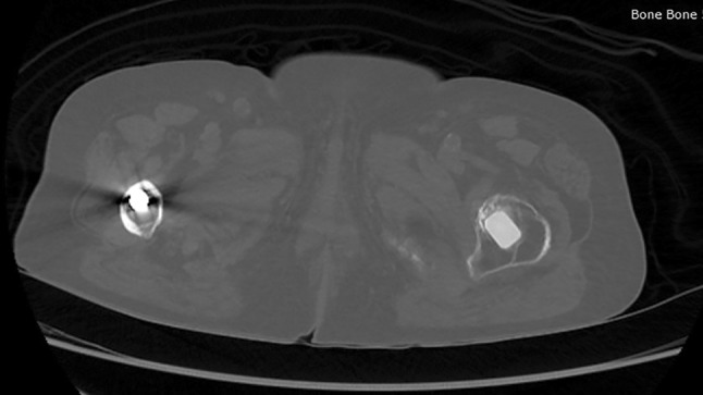
Axial CT section showing the ivory hip stem on the left femur. Please note the high level of osseointegration between the ivory prosthesis and bone after 20 years. An Austin Moore hemiarthroplasty stem can be seen in the right femur.
Official websites use .gov
A
.gov website belongs to an official
government organization in the United States.
Secure .gov websites use HTTPS
A lock (
) or https:// means you've safely
connected to the .gov website. Share sensitive
information only on official, secure websites.
Axial CT section showing the ivory hip stem on the left femur. Please note the high level of osseointegration between the ivory prosthesis and bone after 20 years. An Austin Moore hemiarthroplasty stem can be seen in the right femur.