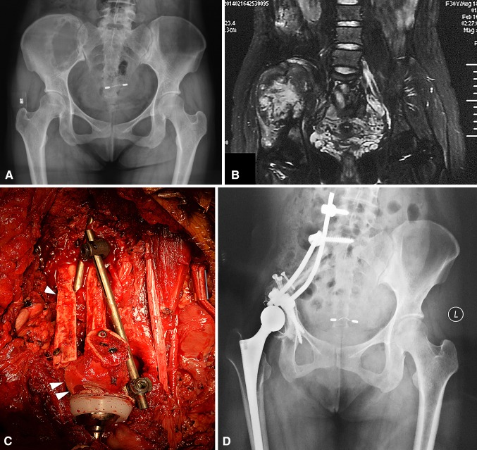
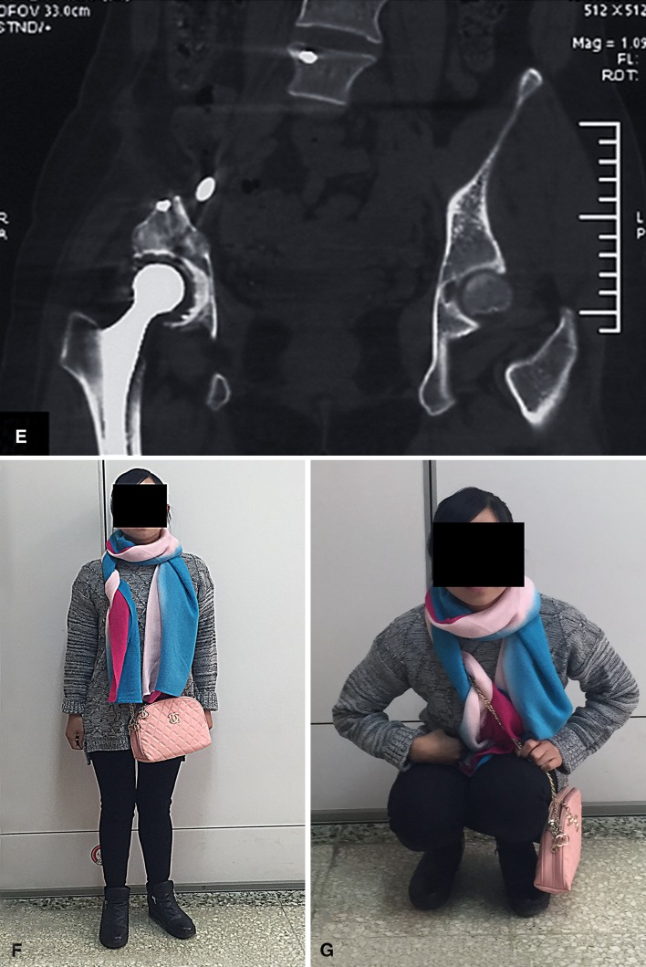
Fig. 3A–H.
A female patient (Patient 10) with pelvic dedifferentiated chondrosarcoma underwent femoral head autograft reconstruction. (A) Her preoperative plain radiograph and (B) T2-weighted MR image show involvement of the ilium and superior part of the acetabulum. (C) An intraoperative view shows the pelvic ring is reconstructed with a double nonvascularized fibular autograft (one arrow) and femoral head autograft (two arrows) enhanced by cancellous compression screws and a spinal pedicle screw-rod system. (D) A postoperative plain radiograph and (E) CT scan obtained at the 24-month followup show good local tumor control with no signs of mechanical failure of the endoprosthesis or internal fixation. (F) This patient could stand and (G) squat freely without aid at her 24-month followup.


